


📞 Horsham :01403 597373
📞 East Grinstead :01342537610
Category ANRC Physiotherapy Clinic
16 Jan
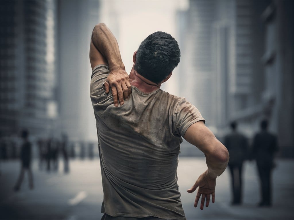
Thoracic Outlet Syndrome (TOS)by Physio Horsham
The term ‘thoracic outlet syndrome’ describes compression of the neurovascular structures as they exit through the thoracic outlet (cervicothoracobrachial region). The thoracic outlet is marked by the anterior scalene muscle anteriorly, the middle scalene posteriorly, and the first rib inferiorly. Certain anatomical abnormalities can be potentially compromising to the thoracic outlet as well. These include the presence of a cervical rib, congenital soft tissue abnormalities, clavicular hypomobility, and functionally acquired anatomical changes. Soft tissue abnormalities may create compression or tension loading of the neurovascular structures found within the thoracic outlet (such as hypertrophy, a broader middle scalene attachment on the 1st rib or fibrous bands that increase the stiffness…). Congenital Factors: Acquired Conditions: Muscular Causes: Clinical symptoms Signs and symptoms of thoracic outlet syndrome vary from patient to patient due to the location of the nerve and/or vessel involvement. Symptoms range from mild pain and sensory changes to limb-threatening complications in severe cases. Patients with thoracic outlet syndrome will most likely present pain anywhere between the neck, face, and occipital region or into the chest, shoulder, and upper extremity and paresthesia in the upper extremity. The patient may also complain of altered or absent sensation, weakness, fatigue, and a feeling of heaviness in the arm and hand. The skin can also be blotchy or discoloured. A different temperature can also be observed. Signs and symptoms are typically worse when the arm is abducted overhead and externally rotated with the head rotated to the same or the opposite side. As a result activities such as overhead throwing, serving a tennis ball, painting a ceiling, driving, or typing may exacerbate symptoms.
READ MORE
16 Jan
Thoracic Hypokyphosis – physio Horsham
Thoracic hyperkyphosis is described as an excessive anteroposterior curvature of the thoracic spine of greater than 40°. Normal kyphosis angles vary between Clinically Relevant Anatomy The thoracic part of the spine natural kyphosis. This thoracic curvature is the result of a slight wedging of the vertebrae. Line of Gravity Etiology There is no widely accepted definition of hyperkyphosis therefore the prevalence of hyperkyphosis is not precisely known. Biomechanical Factors in thoracic hyperkyphosis Clinical Presentation Management Physical management should be considered as a first-line approach. In terms of medications, antiresorptive or bone-building medications are taken by patients with thoracic hyperkyphosis due to their low bone density or spine fractures. Osteoporosis treatment helps to prevent incident spine fractures, however, no medications have been shown to improve hyperkyphosis. Physical therapy Physiotherapy for thoracic hyperkyphosis, including manual therapy, taping and bracing, should be implemented in an early stage and is regularly a first-line treatment. The main goal of any therapy for patients with thoracic hyperkyphosis is to reduce the excessive antero-posterior curvature as well as improve the physical function and decrease the pain. Recognition and treatment of hyperkyphosis could contribute to a reduced risk of falls, fractures, and functional limitations. ANRC PHASES OF REHABILITATION
READ MORE
16 Jan
Sacroiliac Joint Syndrome-by physio horsham
Introduction Sacroiliac joint dysfunction is a term used to describe the pain of the sacroiliac joint (SI joint). It is usually caused by abnormal motion (i.e. hyper- or hypo-mobile) or malalignment of the sacroiliac joint. Sacroiliac joint syndrome is a significant source of pain in 15% to 30% of people with mechanical low back pain. Clinically Relevant Anatomy The sacroiliac joints are located on each side of the spine between the two pelvic bones, which attach to the sacrum. The main function within the pelvic girdle is to provide shock absorption for the spine and to transmit forces between the upper body and the lower limbs. The SI joint experiences forces of shearing, torsion, rotation, and tension. Ambulation is heavily impacted by the SI joint, as this is the only orthopaedic joint connecting the upper body to our lower body. The joint is a relatively stiff synovial joint filled with synovial fluid. The bones of the sacrum and ilium are coated in hyaline cartilage at their articular surfaces with dense fibrous tissue connecting the ilium and the sacrum. SI joints typically only have a few degrees of motion. Etiology Injuries to the sacroiliac joint can occur from various etiologies. Epidemiology Pregnancy Approximately 90% of the population will at some experience or present to the clinic with some form of low back pain/pathology. About 10% to 25% of these patients are thought to be experiencing pain from the SI joint. The majority of SI joint pathologies affect the adult patient population. Clinical Presentation Symptoms of sacroiliac joint syndrome are often difficult to distinguish from other types of low back pain. This is why making a diagnosis of sacroiliac joint syndrome is very difficult. The most common symptoms include: FOR FURTHER INFORMATION see your local Physiotherapist. Click here to find your closest ANRC Physio clinic FOR MORE INFORMATION see your local Physio Practitioner. Click here to find your Physiotherapy clinic
READ MORE
16 Jan
How Posture Affects Your Health and Well-Being: A Guide to Understanding and Improving Your Body Alignment
What is Posture? Posture is the attitude assumed by the body either with support during muscular inactivity, or the coordinated action of many muscles working to maintain stability. It forms an essential basis that is being adapted constantly. Our posture is an active process involving not only our muscles and joints but also our perception, emotions, and the environment we are in. Even seemingly static positions, like sitting or standing, are full of tiny adjustments and movements. Posture is a highly individual and dynamic aspect of human physiology. It is more about how your body adapts and interacts with different situations than a fixed ‘correct’ or ‘incorrect’ state. Posture can be simply defined as the way in which we hold our bodies while standing, sitting, or lying down. Inactive Posture: Describe postures adopted during resting or sleeping, and they are more suitable for this purpose when all the essential muscular activity required to maintain life is reduced to a minimum. Active Posture: The integrated action of many muscles is required to maintain active postures, which may be either static or dynamic. Posture and Health Poor posture can be bad for your health. Maintaining a posture that puts stress on a joint such as prolonged slouching (see image at R) or slumping over can: Important advice could include: FOR FURTHER INFORMATION see your local Physiotherapist. Click here to find your closest ANRC Physio clinic FOR MORE INFORMATION see your local Physio Practitioner. Click here to find your Physiotherapy clinic
READ MORE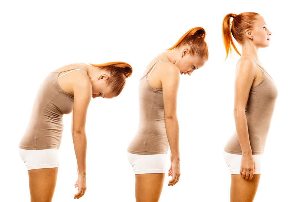
16 Jan
POSTURAL DISORDERS
1. FORWARD HEAD POSTURE The National Academy of Sports Medicine (NASM) defines FHP as holding the head out, in front of its natural position over the cervical spine. A person with FHP also typically tilts their head back in order to look forward. This posture puts a strain on the muscles and bones of the neck. It can also lead to muscle imbalances, as some muscles support more of the load than others. The forward position of the head puts increasing amounts of weight pressure on the spine. The muscles that FHP weakens and lengthens include: The muscles that become shortened and overactive include: Causes People may associate FHP with using electronic devices for a long time, such as cell phones or computers. However, any activity that causes a person to lean their head forward for a prolonged period of time can lead to chronic FHP. Some potential causes of FHP include: Side effects There are several potential side effects and symptoms associated with FHP. Pain and stiffness The extra pressure on the neck muscles can cause increased strain and pressure. This in turn can cause pain and stiffness in the neck muscles. People may experience: Rounded shoulders and upper back FHP can cause a person to develop rounded shoulders and upper back. Additionally, it can impact the movement patterns for the scapula, or shoulder blade, and the humerus, which is the long arm bone, resulting in a condition called scapular dyskinesis. If a person experiences FHP and rounding of the shoulders, healthcare professionals refer to this as upper crossed syndrome. RESPIRATORY ISSUES FHP can cause the upper chest to expand while the lower chest contracts. This change in shape can interfere with regular breathing. Treatment A doctor may recommend physical therapy. There are several potential treatments that a person can try at home, along with medical interventions. A person who works using a computer or other devices can practice sitting upright and keeping their neck in a neutral spine position. This means ensuring that the ears are in line with the shoulders. Poor posture can lead to postural kyphosis. If this is severe, doctors may recommend surgical intervention. In some cases, a person may find that pain medications may be helpful in reducing pain. A person may want to speak with their doctor about muscle relaxants or prescription-strength medications for severe pain. ANRC Phases of Rehabilitation Phase 1- Pain Relief (Trigger Point Therapy, Myofascial Release, Taping, Electrotherapy modalities(ultrasound, tens, IFT), breathing exercises, Basic Home exercises, etc. Phase 2- Restoration of Range of movement of the neck, postural correction by various types of stretching exercises to tighten muscles and strengthening for the weakened muscles, scapular setting exercises with the resistive band, chin tucking exercises to improve the neck flexors strength along with breathing exercise. Phase 3- Maintenance Phase & Improve the activity of daily living (Intensive Strength Training, Ergonomic Modifications in workplaces & Daily Activities), finally concentrating on the core strengthening exercises to make u all fit far better than before, proper advising to the clients about what to do and how to do for further prevention. And from ANRC REHABILITATION CENTRE HORSHAM here we are listing some of the preventive measures that u all can do easily with your daily activities. FOR FURTHER INFORMATION see your local Physiotherapist. Click here to find your closest ANRC Physio clinic FOR MORE INFORMATION see your local Physio Practitioner. Click here to find your Physiotherapy clinic
READ MORE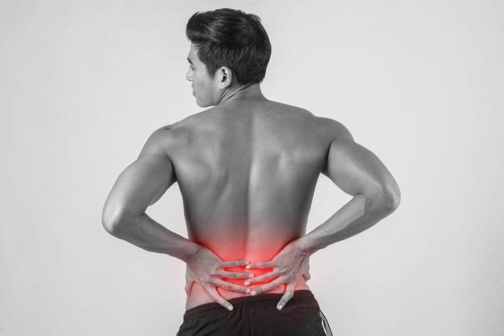
16 Jan
How to Prevent and Treat Low Back Pain Caused by Hyper Lordosis: A Complete Guide to Understanding the Causes, Symptoms, and Solutions for This Common Spinal Condition -Physio Horsham
How to Prevent and Treat Low Back Pain Caused by Hyper Lordosis: A Complete Guide to Understanding the Causes, Symptoms, and Solutions for This Common Spinal Condition -Physio Horsham
READ MORE
19 Apr
Heel Pain (Plantar fasciitis)
Heel Pain (Plantar Fasciitis) Introduction Heel pain is a very common problem that may be attributed to several etiologies, including heel spurs, plantar fascia irritation (acute or chronic), and bursitis. Plantar fasciitis is thought to result from microtears in the fascia due to repeated biomechanical stress on the arch of the foot on weight-bearing. Plantar fasciitis is an inflammation of the plantar fascia & the perifascial structures. Chronic stress to the origin of this fascia on the calcaneus may cause calcium to deposit, forming a spur (plantar calcaneal spur). Plantar Fascia is a Dense, broad band of connective tissue attaching proximal and medially to the calcaneus and fans out over the plantar aspect of the foot and works in maintaining the stability of the foot and bracing the longitudinal arch. The pain is maximal when the patient first stands in the morning and tends to decrease with walking. Ten percent of people may experience pain under the heel (plantar heel pain) at some time during their life. Cause Plantar fasciitis is the most common cause of heel pain in the adult population with an incidence of around 10%. It affects adults across the age spectrum with an incidence in women twice that of men. Ethnicity does not influence incidence. There is a higher preponderance in those who play sport either recreationally or professionally. Increased body weight and an increased body mass index (BMI) have also been shown to be significant risk factors for developing plantar fasciitis. Pregnancy, flat feet, high arched feet, poorly fitting or worn footwear, Calf muscle tightness, gait abnormalities, prolonged standing, running, jumping and walking are additional contributory factors. Excessive lumbar lordosis—a condition in which an increased forward tilt of the pelvis produces an unfavourable angle of foot strike when there is a considerable force exerted on the ball of the foot—can also contribute to this problem. Running on soft surfaces are also potential causes of plantar fasciitis. Clinical features Assessment Looking for Swelling, Arches, Deformity, Gait, posture, tenderness, Tight Structures etc & History Summary For the treatment of heel pain or plantar fasciitis, research has not indicated any consensus on a specific treatment regimen that has proven to resolve heel pain with any statistical significance. However nonsurgical treatment is ultimately effective in approximately 90% of patients. It is equally important to correct the problems that place individuals at risk for plantar fasciitis, such as the increased amount of weight-bearing activity, increased intensity of activity, hard walking/running surfaces and worn shoes. Early recognition and treatment usually lead to a shorter course of treatment as well as an increased probability of success with conservative treatment measures S.M.A.R.T. approach to prevent plantar fasciitis The Canadian Physiotherapy Association (CPA) Calf and Achilles stretching is achieved by performing asymmetrical stretching exercises ANRC Phases of Rehabilitation Phase 1- Pain Relief (Trigger Point Therapy, Myofascial Release, Sports massage, Taping, Electrotherapy modalities, Postural Correction, Basic Home exercises etc) Phase 2- Restoration of Range of movement and muscle length (Stretches, Exercises for the small muscles of the foot, Postural training, Basic Strength Training, Orthotics if needed) Phase 3- Maintenance Phase & Improve the activity of daily living (Intensive Strength Training, Ergonomic Modifications in Sports & Daily Activities Reference
READ MORE
6 Apr
Myofascial Pain Syndrome
Musculoskeletal disorders are the main cause of disability in the working-age population and are among the leading causes of disability in other age groups. Myofascial pain syndrome is a common painful muscle disorder caused by myofascial trigger points. Pain and limited function of the locomotor (or musculoskeletal) system are two of the most common reasons for consulting a doctor or therapist. The fascia & muscles have a key role in this, because of their anatomical and functional properties. The importance of the muscles is frequently underestimated in practice, however, although muscular imbalances, muscle tension and painful disorders of muscle function play a large part in both acute and chronic locomotor system symptoms, according to current knowledge. The clinical correlate is the myofascial trigger point (mTrP). We rely for the most part on the guidelines of the pioneers in the field of trigger point research, Janet Travell and David Simons (Travell, Simons 2002). Simons et al. (1999) have claimed that myofascial trigger points (TrPs) from neck and shoulder muscles might play an important role in the genesis of mechanical neck pain. There are epidemiological studies suggesting that TrPs represent an important source of musculoskeletal disorder (Chaiamnuay et al., 1998). We understand the mTrP to be a site or band that is hypersensitive and palpably tense compared to the surrounding area in a muscle that is often shortened, and which demonstrates changes in tone and consistency (‘taut band’). It is painful when palpated and from it pain and autonomic disorders may be caused in an area that cannot usually be attributed to a particular segment (‘referred pain’). We describe the resulting muscle pain as myofascial pain syndrome (MPS). The formation of TrPs may result from a variety of factors, such as severe trauma, overuse, mechanical overload or psychological stress (Simons et al., 1999). Recent studies have hypothesized that the pathogenesis of TrPs results from injured or overloaded muscle fibres. This leads to involuntary shortening, loss of oxygen supply, loss of nutrient supply and increased metabolic demand on local tissues (Mense et al., 2000). Diagnostic criteria are: A localised, dull, pressing, dragging, occasionally burning spontaneous pain associated with acute or chronic muscular strain The pain is often described as spreading or radiating. Referred pain is an important characteristic of a trigger point Tenderness with typical pain reproduction within a palpable ‘taut band’ of muscle, A pain that predominantly radiates in a distal direction after mechanical stimulation, Painful limitation of movement, Muscular weakness without atrophy. As these are not ‘hard’, evidence-based criteria but subjective information and findings, an appropriate finding oriented medical differential diagnosis is an indispensable prerequisite for making the diagnosis of ‘myofascial pain’ with or without limitation of movement In the head and neck region, myofascial pain syndrome with trigger points can manifest as tension headache, tinnitus, temporomandibular joint pain, eye symptoms, and torticollis. Upper limb pain is often referred and pain in the shoulders may resemble visceral pain or mimic tendonitis and bursitis. In the lower extremities, trigger points may involve pain in the quadriceps and calf muscles and may lead to a limited range of motion in the knee and ankle. The muscular fascial network suggests the following applications in relation to trigger points: pain sensations caused by trigger points supposedly originate mostly from fascial receptors which have undergone sensitisation. A possible treatment plan is a therapeutic stimulation and proprioceptive sensitisation of other fascial receptors (preferably in the area belonging to the same cortical area). Fascial changes supposedly also play a key role in changed muscle stiffness in the area around a trigger point as well. Principles of Treatment: PHYSIOTHERAPIST –PATIENT RELATIONSHIP: The key influential factor for the success of treatment is a ‘healing’ physiotherapist–patient relationship. In patients with chronic myofascial pain, in particular, psychosocial stress factors must also be evaluated and included early on in the consultation. The treatment plan for myofascial trigger point therapy (MANUAL TECHNIQUES AND DRY NEEDLING) – Ischaemic compression of the mTrP, Manual stretching of the mTrP region Fascia stretching technique, Manual stretching of the superficial and intramuscular fascia etc Functional training, ergonomics, Physiological weight-bearing and exercise support the regeneration process and make the muscles more resilient Ergonomics reduces incorrect strain on the muscles Summary The aim of trigger point therapy is to provide a permanent cure for myofascially caused pain and functional disorders. Otherwise, recurrences inevitable This requires: finding the relevant active trigger point, deactivating the relevant trigger point using manual techniques and/or dry needling, providing thorough manual treatment of the connective tissue changes which occur as a reaction, especially with chronic symptoms, recognising the factors which cause the problem to persist and including them in the treatment process Predisposing and perpetuating factors must be recognised and included in the treatment strategy in the form of ergonomics and functional training of the muscles ANRC Phases of Rehabilitation Deactivation of trigger points and Pain Relief ( Trigger Point Therapy, Myofascial Release, Taping, Electrotherapy modalities, relaxation Training etc) Restoration of Range of movement – Stretches, Postural training, Basic Strength Training Maintenance Phase & Improve the activity of daily living – Intensive Strength Training, Ergonomic Training etc Reference Myofascial Trigger Points Comprehensive diagnosis and treatment; Edited by Priv. Doz. Dr. med. Dominik Irnich: Toronto 2013 First edition published in English © 2013, Elsevier Limited. All rights reserved. Trigger Points: Diagnosis and Management DAVID J. ALVAREZ, D.O., and PAMELA G. ROCKWELL, D.O., University of Michigan Medical School, Ann Arbor, Michigan. Myofascial trigger points in subjects presenting with mechanical neck pain: A blinded, controlled study C. Ferna´ ndez-de-las-Pen˜ as_, C. Alonso-Blanco, J.C. Miangolarra Department of Physical Therapy, Occupational Therapy, Physical Medicine and Rehabilitation, Universidad Rey Juan Carlos (URJC), Alcorco´n, Spain Received 12 November 2004; received in revised form 4 January 2006; accepted 3 February 2006
READ MORE
10 Mar
What is Interferential Current Therapy ?
Interferential Current Therapy Interferential current therapy is an effective therapy option used by many physiotherapy clinics to relieve pain and accelerate the self-healing process, getting your body back to a healthy, pain-free state. The high-frequency signals of an IFC penetrate through the skin into deeper lying muscle tissues. Electrodes are placed on your skin around the injured body part. The Interferential Current device then transmits electrical impulses in minute quantities through your skin. Underlying tissue and nerves are stimulated which begins the healing properties. These impulses are not painful in the least. In fact, patients describe the sensation as a minor prickle on their skin. Frequencies produced by the IFC have been proven to stimulate endorphins, the body’s natural pain killers. This can help to create a self-healing process without the need to for medications. This form of therapy is also extremely useful in reducing pain, inflammation, curing edema, and spasms. Reduces or eliminates your pain safely. Noticeable decrease in swelling and inf Restores lost movement and improve restricted movements and coordination. Stimulates the natural hormones which can help your body heal faster. Considered by many experts as a highly effective form of treatment for chronic pain. How the Interferential Current Therapy Helps Interferential Current stimulation is very useful in the treatment of circulatory and muscular disorders, stiffness of joints, edema, and inflammation. If you suffer from health problems such as cumulative trauma disorders, body pain, joint injuries, or are pre or post orthopedic surgery, interferential current therapy is an important option. Interferential current therapy has been in use for many years, and there have been numerous case studies and research reports that have documented its versatility in treating diverse symptoms, accelerating the healing process and restoring normal movement. Patients who chose to undergo interferential current therapy have fewer post-op complications compared to people who rely exclusively on medications for pain relief. It also helps in blood circulation and hastens the healing process by stimulating endorphins. Common Questions Are interferential devices safe? Does my insurance cover the cost for interferential treatment? Where does interferential currency therapy work best? Is interferential current therapy better than TENS? How does interferential current work? Coverage Options ANRC physiotherapy services are covered by most health insurance companies. Not sure if you’re covered? No problem. We can help you find out (and usually within the hour). Just call us at Hotline: 07483 807551 Horsham: 01403 597373 East Grinstead/Lingfield: 01342 537610 At ANRC we believe your health should come first. We take care of the paperwork so you can focus on getting better, plus we offer direct insurance billing, saving you time and upfront costs. Learn More About pt Health Coverage Options Research The journal physical Therapy published a research summary in 2006 which talked about interferential therapy used in various models of inflammatory pain. TheIrish Medical Journal published a research summary in 1989 which discussed “interferential therapy for the treatment of stress and urge inconsistency”.
READ MORE
17 Oct
Importance of pelvic floor strengthening exercises re-emphasised
UK physiotherapists used Valentine’s Day to launch a campaign to encourage members of the public to perform physiotherapy exercises to tighten up their pelvic floors. Pelvic floor muscle exercises tone and strengthen these muscles, improving blood supply and nerve activity, all leading to greater pleasure. Many people are already aware of the importance of tightening the Kegel muscles – those used to stop urine mid-flow – in order to tighten the pelvic floor, but the campaign highlights that it’s also important to practice tightening the muscles around the anal sphincter too. Studies have shown that doing this can assist in bringing the vaginal walls together for women, while it can also help men to sustain an erection. In addition, the campaign emphasises the importance of relaxing the muscles after each contraction and suggests fitting in these exercises while performing other activities, such as brushing teeth. Experts recommend squeezing these muscles ten to 15 times in a row at least once a day and that each squeeze should be short and powerful. Varying the strength of contractions is also important; physios suggest trying to maintain a squeeze for five to ten seconds regularly too, while continuing to breathe normally. (Article from Just Physio)
READ MOREQuick Contacts
- Phone : +44 7483 807551
- Email : info@anrc-uk.com
- Locations : Horsham, East Grinstead, Ashurstwood, Lingfield
- Country : United Kingdom


